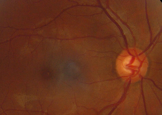
Physiology of Optic Disc
The optic disc, also known as the optic nerve head, is the point at which the optic nerve fibers leave the eye and enter the brain. The physiology of the optic disc is critical in understanding the normal functioning of the visual system and the causes and effects of eye diseases.
Function of the Optic Disc
The main function of the optic disc is to transmit visual information from the retina to the brain. The retina is the light-sensitive layer of tissue at the back of the eye that converts light into electrical signals. These electrical signals are then transmitted to the brain via the optic nerve. The optic disc is where the optic nerve fibers converge to form the optic nerve.
The optic disc also plays an important role in maintaining the health of the retina. The blood vessels that supply the retina with blood and oxygen enter and exit the eye through the optic disc. The optic disc also contains a small number of retinal pigment epithelial cells that help to protect and support the retina.
Structure of the Optic Disc
The optic disc has a central depression called the optic cup. The optic cup is where the nerve fibers converge to form the optic nerve. The rim of the optic disc is called the neuroretinal rim, and it is made up of retinal ganglion cells and their axons. The optic disc is surrounded by the peripapillary sclera, which is the white part of the eye. The peripapillary sclera is thicker and more fibrous than the sclera in other parts of the eye, providing additional support for the optic disc.
Blood Supply of the Optic Disc
The optic disc is supplied with blood by the central retinal artery and vein. The central retinal artery enters the eye through the optic disc and branches out to supply blood to the retina. The central retinal vein exits the eye through the optic disc, carrying blood away from the retina.
Pathology of the Optic Disc
Damage to the optic disc can result from a variety of eye conditions, including glaucoma, diabetic retinopathy, and age-related macular degeneration. These conditions can cause changes in the appearance of the optic disc, such as a thinning of the neuroretinal rim or an enlargement of the optic cup. These changes can be detected through a comprehensive eye examination, including a dilated fundus examination, and can help to diagnose and monitor these conditions.
Conclusion
The physiology of the optic disc is critical in understanding the normal functioning of the visual system and the causes and effects of eye diseases. The main function of the optic disc is to transmit visual information from the retina to the brain. The structure of the optic disc includes a central depression called the optic cup, the neuroretinal rim and the peripapillary sclera. The blood supply of the optic disc is through the central retinal artery and vein. Damage to the optic disc can result from a variety of eye conditions, which can be diagnosed and monitored through a comprehensive eye examination.

Comments
Post a Comment